Introduction
When it comes to diagnosing certain medical conditions, accurate and targeted sampling of tissues or cells is of paramount importance. CT (Computed Tomography) and USG (Ultrasonography) Guided FNAC (Fine Needle Aspiration Cytology) are two remarkable procedures that have revolutionized tissue sampling, providing doctors with invaluable insights for diagnosis. Let’s embark on a journey into the world of CT and USG Guided FNAC and explore how they make the diagnostic process more precise and efficient with easy-to-understand language.
What is CT & USG Guided FNAC?
CT and USG Guided FNAC are advanced imaging-guided procedures used to extract samples of tissue or cells from specific areas of the body for further examination under a microscope. These procedures help doctors diagnose a wide range of medical conditions, including tumors, cysts, and infections, without the need for invasive surgery.

How does it work?
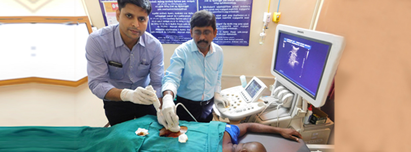
During CT or USG Guided FNAC, the patient lies on a table, and the CT or ultrasound machine is used to precisely locate the target area. A thin needle is then inserted through the skin into the targeted tissue or lesion to obtain a small sample. The procedure is performed under local anesthesia, ensuring minimal discomfort for the patient.
The collected sample is then sent to a pathology laboratory, where it is examined under a microscope by a pathologist to make a definitive diagnosis.
Advantages of CT & USG Guided FNAC
These imaging-guided FNAC procedures offer several key benefits:
- Precision: CT and USG provide real-time imaging, allowing doctors to precisely target the area of interest, ensuring accurate sample collection.
- Minimally Invasive: Since only a thin needle is used, there is no need for major incisions or surgery, resulting in less pain, reduced risk of complications, and faster recovery.
- Speedy Results: The collected samples are quickly sent to the pathology lab for analysis, providing doctors with prompt results, allowing for timely treatment decisions.
- Safe and Effective: CT and USG are well-established imaging techniques that are considered safe and effective for guiding FNAC procedures.

Applications of CT & USG Guided FNAC
These imaging-guided FNAC procedures find applications in various medical fields, including:
- Oncology: CT and USG Guided FNAC are commonly used to diagnose and stage cancers, helping doctors plan the most appropriate treatment strategies.
- Thyroid Nodules: FNAC guided by ultrasound is often employed to assess thyroid nodules and determine whether they are benign or cancerous.
- Lung Lesions: For suspicious lung lesions detected on CT scans, CT-guided FNAC aids in making a precise diagnosis.
- Abdominal Masses: USG-guided FNAC is valuable in diagnosing abdominal masses, such as liver or kidney tumors.
Conclusion
CT and USG Guided FNAC have transformed the way tissue and cell samples are obtained for diagnosis. These minimally invasive and highly precise procedures have made it easier for doctors to identify and treat various medical conditions promptly and accurately.
As technology continues to evolve, we can expect further improvements in imaging-guided procedures, making medical diagnosis and treatment even more precise and patient-friendly. Until then, we can marvel at the wonders of CT and USG Guided FNAC – navigating the path to a precise diagnosis with ease.
