Introduction
In the realm of medical diagnostics, X-rays have been an essential tool for many decades. They have allowed doctors to see inside the human body without the need for surgery, providing valuable insights into various medical conditions. But what happens when technology meets X-rays? That’s where Digital X-Ray comes in – a revolutionary advancement that has transformed the way we capture and interpret X-ray images.
Let’s explore the world of Digital X-Ray and understand how it has made a significant impact on medical imaging with simple and easy-to-understand language.
What is Digital X-Ray?
Digital X-Ray is a modern technique that replaces traditional X-ray films with digital sensors to capture images of the body’s internal structures. Instead of using film to record the X-ray image, digital detectors instantly capture the data and convert it into high-quality digital images that can be viewed on a computer screen.

How does it work?
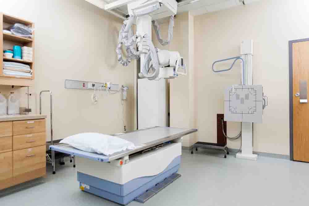
The process of obtaining a Digital X-Ray is quite similar to traditional X-rays, but with a notable difference. The patient is positioned between the X-ray machine and the digital detector, and the X-rays pass through the body to create an image.
However, instead of developing the image on a film, the digital detector immediately sends the X-ray data to a computer. Powerful software processes this data and generates detailed images of the body’s bones, organs, and tissues.
Advantages of Digital X-Ray
Digital X-Ray brings a host of benefits that have significantly improved medical imaging:
- Faster Results: With traditional X-rays, the film needed to be processed, which could take some time. Digital X-Ray provides instant results, allowing doctors to view and analyze images immediately. This speed is particularly crucial in emergency situations.
- Enhanced Image Quality: Digital X-Ray produces clearer and more detailed images compared to traditional X-ray films. The higher resolution allows doctors to see even small abnormalities, leading to more accurate diagnoses.
- Reduced Radiation Exposure: Digital X-Ray requires less radiation to produce images of comparable quality to traditional X-rays. This means patients are exposed to lower levels of radiation, enhancing their safety during the procedure.
- Easy Storage and Sharing: Digital X-Ray images can be stored electronically, eliminating the need for physical storage space for X-ray films. Furthermore, these images can be easily shared with other healthcare providers, making it convenient for consultations and second opinions.
- Image Enhancement: The digital nature of these images allows for easy manipulation and enhancement. Doctors can zoom in, adjust brightness and contrast, or even annotate specific areas for better visualization and communication.


Applications of Digital X-Ray
Digital X-Ray is used across various medical fields, including:
- Orthopedics: It is instrumental in diagnosing bone fractures, joint problems, and spine conditions.
- Dentistry: Digital dental X-rays aid in detecting dental problems, like cavities and tooth decay.
- Chest X-Rays: It is valuable in diagnosing lung conditions, such as pneumonia and tuberculosis.
- Trauma Cases: Digital X-Ray quickly assesses injuries after accidents, helping guide immediate treatment.
Conclusion
Digital X-Ray has undoubtedly revolutionized medical imaging, allowing doctors to obtain clearer and more immediate insights into the human body. The advantages it brings, including faster results, enhanced image quality, and reduced radiation exposure, have made it a vital tool in modern healthcare.
As technology continues to advance, we can expect even more improvements in the field of medical imaging. Until then, we can appreciate the wonders of Digital X-Ray – a remarkable marriage of technology and medicine that benefits patients and healthcare providers alike.
